In my last post I gave an introduction and brief description of the Microflex that I bought on ebay. Since then I’ve had the opportunity to try it out on a few stained specimens to see how well it performs.
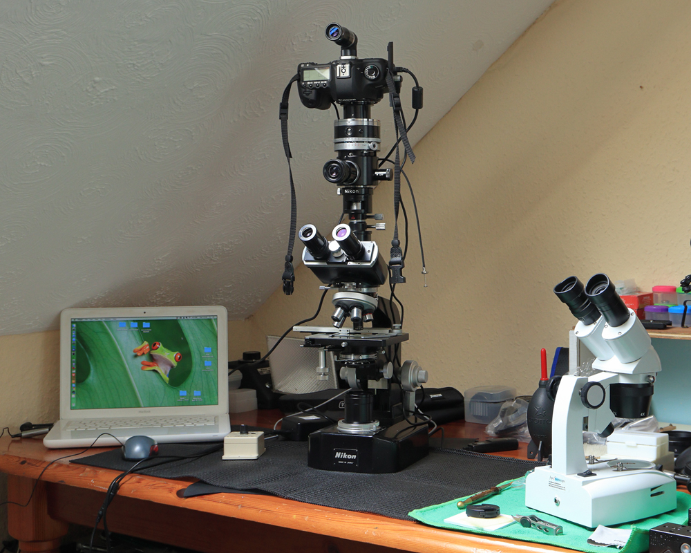
I started using the Microflex as it was intended to be used in the early 1970s i.e. without the use of a computer. It’s a precision piece of engineering and looks great on my Nikon K-Le microscope. Here is a breakdown of my initial working procedure:
Method 1
- Turn the dial on the Microflex body to the desired shutter speed – 1-1/250 sec. (manual, metered); 32-2 sec., B, T (manual). I use the B or T setting in order to keep the shutter blades open and allow the camera’s exposure meter to decide which shutter speed to use.
- Focus the specimen through the right-angle finder using Kohler illumination. The finder gives a bright and parfocal focus to the camera sensor and composing is made easier by placing the image within the Full-frame or APS-C frames, etched onto the viewfinder screen.
- Use mirror lock-up and press the shutter release. The mirror in the camera flips up.
- Wait approximately two seconds for the mirror vibrations to vanish and fire the shutter a second time. The sensor has now been exposed to the image.

Aphid – 16 image stack using Zerene Stacker
Vintage Nikon L-Ke microscope
Nikon 4x Plan 0.1 objective and Nikon 5x eyepiece
total magnification of x20
Method 2
Mainly as above but using the Live View function on the camera
- Similar to method 1 but using Live View. The camera is set to silent mode in order to activate an electronic shutter instead of the normal mechanical one. This is much better for high magnification work as it reduces any chance of vibration through the opening of the shutter mechanism.
- Instead of using the right-angle finder, focusing is done through the camera LCD screen.
- In Live View mode the mirror is automatically locked up so I only need to press the shutter release once.
- The shutter fires and is noticeably quieter than method 1.

Nikon 2.5x Plan 0.065 objective and Nikon 5x eyepiece
giving a total magnification of 12.5x.
As expected the 18 image stack clearly has more detail. Both images received the same exposure of 1/13 / ASA 100, however the 18 stack is clearly darker and appears over exposed. Perhaps when stacking I should increase the exposure. Will need to experiment further with stacking software.
Method 3
This is my preferred method as it uses most of the procedures of method 1 but with the added benefit of using the electronic shutter of method 2. It also has the benefit of being able to use a computer screen to greatly aid with focusing.
- Comparable to method 2 but with the camera connected to a laptop via USB lead.
- Canon EOS Utility is then used to control the camera remotely.
- The right-angle finder can still be used with the course focus knob for initial focus. The focus can then be enlarged on the laptop monitor for fine focus tweeking.
- In Live View mode the mirror is automatically locked up so I only need to press the shutter release on the EOS Utility control panel.
- Apart from the focusing (which must be done through the course and fine focus knobs on the microscope) most camera settings are set through the control panel. This greatly reduces the risk of any vibration caused by accidently moving or touching any camera or microscope while the shutter is released.
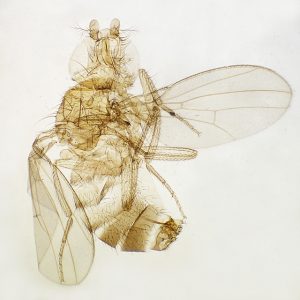
Fruit Fly stained specimen – Single shot.
Vintage Nikon L-Ke microscope – Nikon 4x Plan 0.1 objective
and Nikon 5x eyepiece giving a total magnification of 20x
Pros:
- Beautifully crafted piece of engineering that compliments the classic look of the vintage Nikon S series microscopes.
- Solid and built like a tank. The fact that it is heavy may be seen as a negative point but for me it’s a definite positive. Its weight will help to dampen out any vibrations caused by mirror and shutter activation. If you doubt this then consider the use of a heavy macro rail or tripod as opposed to a light one. A heavy apparatus provides a far more solid and stable support for a camera and hence less likely to cause any unwanted movement.
- As the name Microflex suggests, it can easily be adapted to different ways of working, with or without LiveView; with or without a computer.
- The right-angled viewfinder is parfocal (what you see in focus on the viewfinder is also in focus on the sensor). The Full-frame and APS-C frames on the viewfinder are also useful for framing the specimen.
- Excellent value for money. Not easy to find but some great bargains to be found on ebay. Classic Microflex systems of the 1960s/70s were classy and wonderfully engineered units and worth many times more than the asking price.
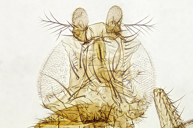
Fruit Fly stained specimen
10 image stack using Zerene Stacker – Vintage Nikon L-Ke microscope
Nikon 10x Plan 0.25 objective and Nikon 5x eyepiece
giving a total magnification of 50x
Cons:
- It’s almost 50 years old, so if using the leaf shutter, don’t expect the speeds to be accurate. Use the camera electronic shutter.
- Adds considerable height to the microscopes trinocular tube.
- Some may consider it over engineered and complex.
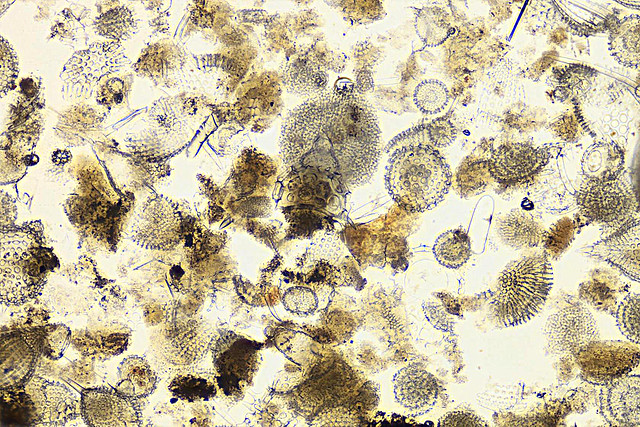
The Radiolaria, also called Radiozoa, are protozoa of diameter 0.1–0.2 mm that produce intricate mineral skeletons, typically with a central capsule dividing the cell into the inner and outer portions of endoplasm and ectoplasm. The elaborate mineral skeleton is usually made of silica.
14 image stack using Zerene Stacker – Vintage Nikon L-Ke microscope
Nikon 10x Plan 0.25 objective and Nikon 5x eyepiece
giving a total magnification of 50x
Conclusion:
The quality of the images are mainly due to the objectives used. In order to avoid chromatic aberration I use Vintage Nikon 10x or 5x eyepieces and plan objectives. These were all made in the 1960/70s and were all designed to be used together. On the whole, I’m pretty pleased with the quality of the images and they’re certainly an improvement over my last set-up. The images can certainly be improved and with further experimentation I’m sure I can increase their definition and sharpness.
What impresses me most is the quality of the Microflex. It’s a fitting testament to the mechanical/engineering excellence of the1960s/70s. The Microflex, are old 1970s pieces of equipment that are beautifully crafted and still give excellent performance almost 50 years later.
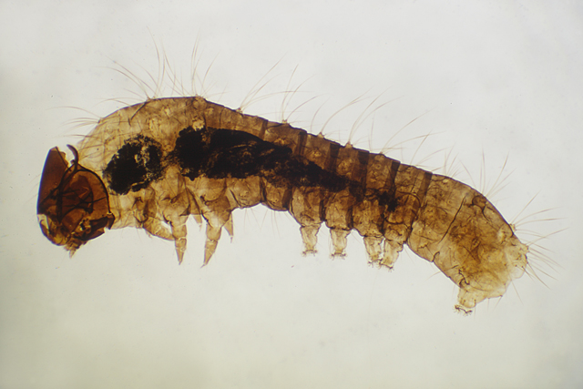
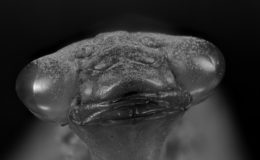
Silk Worm stained specimen – Single shot.
Vintage Nikon L-Ke microscope – Nikon 4x Plan 0.1 objective
and Nikon 5x eyepiece giving a total magnification of 20x

Leave a Comment