In contrast to the all pervading good force summoned upon by Jedi Masters such as Yoda, Obi-Wan Kenobi or the young Luke Skywalker; ‘Darkfield’ sounds like an equally pervasive but albeit altogether more sinister force, more likely to be beckoned upon by Darth Vader or the evil Emperor Palpatine. It may come as a bit of an anti climax when I tell you that the ‘Darkfield’ in question is not some super cosmic, powerful consciousness but an illumination technique used in microscopy. Please bear with me and I hope you’ll agree that the beautiful images possible using ‘Darkfield’ have a luminescent other worldly quality that are perhaps more in tune to sci-fi than to the non-fiction genre.
I’m learning
how to use my microscopes and studying for the first time how to really use
them more effectively, so that I can take more pictures of other cool things
than the standard macro lenses. I’ve finally got some prepared specimen slides,
mainly of insects and aquatic life. For this stuff, dark field microscopy (where
you get bright objects against the dark background) provides more dramatic contrast
than the normal ‘brightfield’ view. The advantage is that you can see details
that are normally not resolved by the microscope objective. An interesting and
perceptive analogy to ‘dust in a room’ was made by Mike Samworth and Wim van
Egmond:
“In a well lit room you do not see the
very small dust particles. However, if the lights go out, a beam of light from
an acute angle makes these same particles visible. Besides the optical
advantages darkfield illumination is very beautiful and gives almost science
fiction like image.”
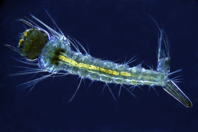
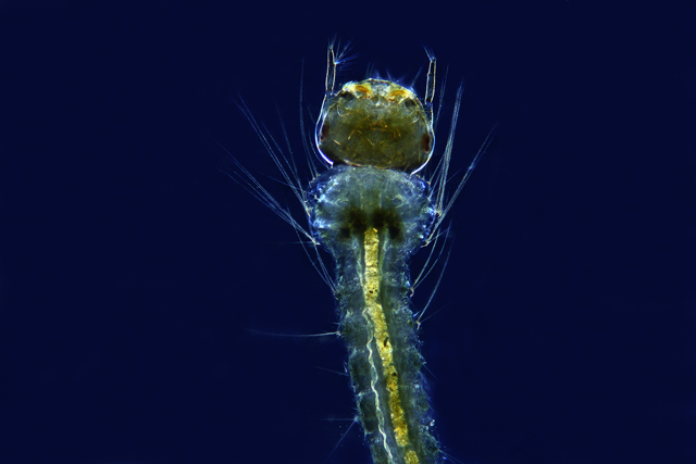
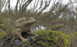
Mosquito Larva – 100x
Bright objects against a dark background provides more dramatic contrast than the normal ‘brightfield’ view.
I used my Nikon L-Ke microscope with phase contrast condenser and 33mm filter holder. However, I didn’t have any “real” patch stops for the filter holder for doing darkfield but was able to experiment just to see how it worked by rotating the phase anulli on the condenser so as to provide oblique illumination and it seemed to partly work OK (100x annuli worked best for this). The main problem appeared to be that the oblique illumination (necessary for darkfield) tended to highlight any dust particles (and these were considerable). A spotless condenser lens would prevent this from happening but that is a tall order when using a vintage 50+ year old microscope!
The next step was to make my own darkfield patch stops and fortunately this was very easy to do myself. What you have to do is place an opaque round stop just below the condensor. An easy way is to cut a piece of black paper and put it on a filter in your filter holder. You can put the stop on a piece of clear acetate sheet. You can even try to draw the stop on it with black paint, place a coin or use a piece of blu tack on the filter. The most important thing is to have the patch big enough to stop all light going directly into the objective. Only the light that is reflected by the objects in the sample reaches the objective.
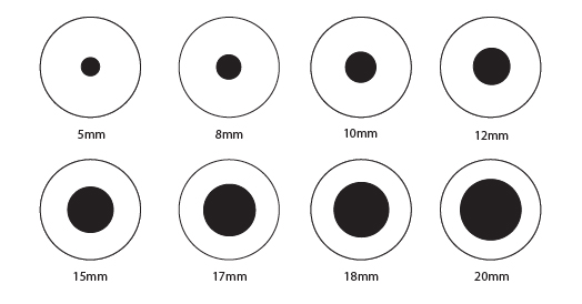
Printed template for patch-stops
Turning to the computer and using Adobe Illustrator I was able to render a series of patch stops that I then printed and used as a template. Once the 33mm acetate circles had been cut out I then had the fiddlier task of cutting out the various patch stops and for this I used sticky backed black velvet. Once the patch stops had been cut out it was simply a case of sticking them to the pre-cut acetate filters.
After some trial and error I found that the ideal stop sizes are 12mm for the 4x objective, 17mm for the 10x objective, and 20mm for the 40x objective. These sizes seem to give the best contrast all around for each objective. However, if anyone is looking for a single solution, the 17mm stop seems adequate for all three objectives (it’s a little dark at 4x, and there’s a little less contrast at 40x, but it’s OK).
One difficulty of darkfield is trying to create a dark field that is truly black. Using the homemade acetate filters with black circular stops sitting just beneath the condenser mainly produced dark blue as opposed to black backgrounds. To keep things simple I used mainly the 10x objective but no matter what sized stopper I used I could not achieve a jet-black background. I suspect this may have been due to getting some direct light creeping past the stop into the objective. Perhaps raising the stop, will effectively make it larger, relative to the aperture of the objective ensuring that the only light entering the system, is outside the aperture of the objective and thus ensuring a darker background. This is something that I will need to experiment with further.
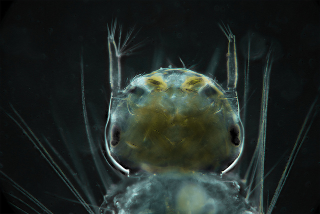
Mosquito Larva 40X
One difficulty of darkfield is trying to create a dark field that is truly black.
Another problem I encountered consisted of capturing backgrounds that were full of specks and dust, no doubt due to imperfections within the slides/cover slips. These imperfections occur with mounts that are thick or when too many light-scattering contaminating artifacts are captured in the mounting medium. Other than have the specimen re-mounted in a thinner mount or mounting medium I was left with the tedious job of having the contaminants removed using the clone tool in Photoshop.
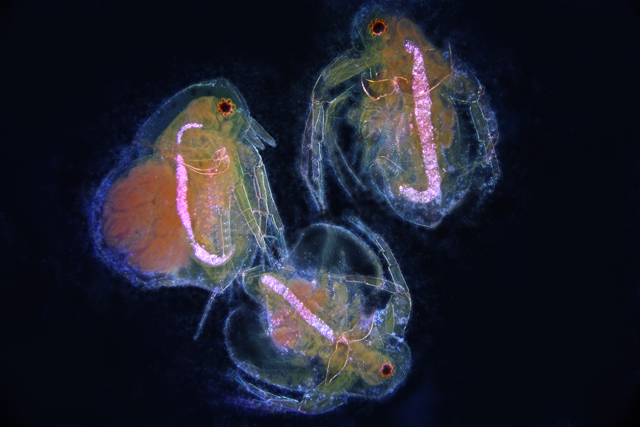
Daphnia – 100x
Before: The background is full of specks and dust, no doubt due to imperfections within the slides/cover slip.
Daphnia, a genus of small planktonic crustaceans, are 0.2–5 millimetres in length. Daphnia are members of the order Cladocera, and are one of the several small aquatic crustaceans commonly called water fleas because their saltatory swimming style resembles the movements of fleas.
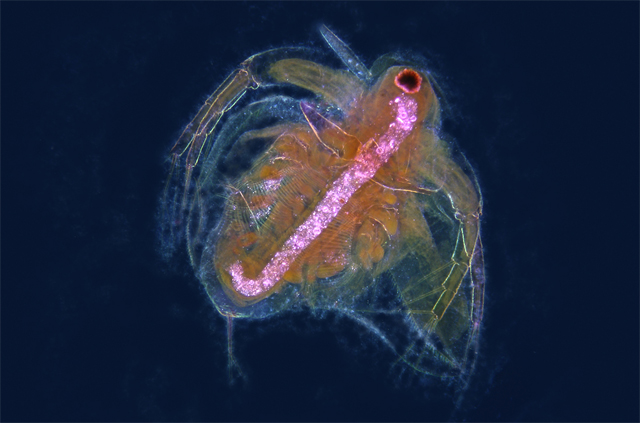
Daphnia – 100x
After: Contaminants removed using the clone tool in Photoshop.
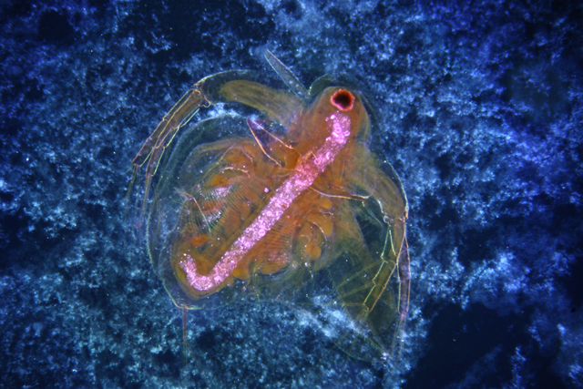
If you look closely you will see every part of its body. Notice the legs that are not used as we would commonly assume to swim but to direct food into the animal’s mouth as well as a stream of water which contains a certain amount of dissolved oxygen without which it could not live. The pink tube running down its body is the gut or alimentary tract and the fluted red circle on its head is the eye.
Daphnia – 40x
Daphnia are interesting to study because we can see their insides, as well as their outside parts.
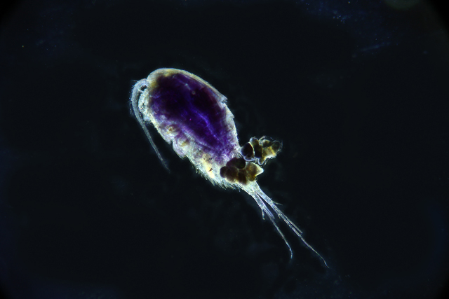
Cyclops – 100x
Cyclops are tiny crustaceans of the family Cyclopidae, also called water fleas.
The two pear-shaped masses attached to the body, one on each side of the tail-like appendage which extends out from the tip of the abdomen are eggs. In time these eggs will hatch but the hatchlings will look nothing like their mother. It is only after they have changed their skin several times that they begin to resemble their parent.
While dark field can create beautiful images under the right circumstances, there are a number of disadvantages to dark field microscopy:
1. Dark field needs an intense amount of light to work. This intense light can create glare and distortion.
2. Dark field is sensitive to contaminants. You need to be meticulous about cleaning your specimen slides and all optical surfaces when performing dark field imaging, as every speck of dirt will want to light up when using dark field. You also need to be careful when preparing your specimens, as any contaminates (dust, air bubbles, etc) in your mounting, above or below the plane of focus, can degrade your image.
3. Specimens, which are not thin enough, are prone to degradation and distortion. The best dark field specimens should be thin to reduce diffraction artifacts.
Leave a Comment